

Dental Filling (Restorative Dentistry): Clinical Indications, Material Selection, and Application Protocols
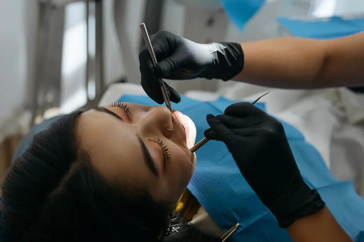
Organic acids released when cariogenic bacteria in the oral flora ferment carbohydrate-containing foods cause mineral loss (demineralization) in the enamel tissue, the outermost layer of the tooth. When this process is not brought under control, enamel integrity is disrupted and the destruction progresses to the dentin tissue in the underlying layer, creating permanent tissue voids called "cavities." In our clinical practice, the procedure of restoring the anatomical form, chewing function, and contact points with adjacent teeth of teeth whose structural integrity has been lost due to decay, wear, or trauma after removing infected tissues is called dental filling (restorative dentistry).
Clinical Diagnostic Criteria and Filling Indications
Not every enamel discoloration or initial cavity requires a filling; enamel lesions that have not yet lost surface integrity (non-cavitated) are monitored with topical fluoride applications and oral hygiene education. However, when destruction reaches the dentin layer and micro-voids form that the patient cannot clean by brushing, restorative intervention becomes necessary. As dentists, in addition to intraoral mirror and probe examination, we make a definitive diagnosis with "bitewing" radiographs, which are especially useful for detecting hidden cavities in the interproximal areas where teeth face each other.
The main conditions for which we indicate fillings in the clinic are as follows:
Primary dental caries that have crossed the enamel and dentin junction and formed cavitation.
Secondary caries that develop under a filling due to loss of marginal adaptation or microleakage in old restorations in the mouth.
Non-carious tissue losses occurring in the cervical (neck) areas of teeth at the gum line due to clenching (abfraction), hard brushing (abrasion), or consumption of acidic foods (erosion).
Enamel-dentin fractures caused by trauma that do not expose the nerve tissue (pulp).
Restorative Materials Used and Their Biomechanical Properties
Amalgam fillings, the mercury-silver alloy used frequently in past years, have been abandoned in our clinical practice because they do not chemically bond to tooth tissue, require removal of sound, non-carious tooth tissue to provide retention, and, due to the metal's coefficient of expansion, can cause cracks in the tooth walls. Today, three main material groups are used according to the biomechanical requirements of the case:
Composite Resin Fillings: These are tooth-colored direct restorative materials composed of inorganic glass filler particles and an organic resin matrix. They bond to tooth tissue micromechanically via "adhesive" (bonding) agents. They do not require removal of healthy tooth tissue for retention; cleaning out the decayed tissue alone is sufficient. They show high resistance to chewing forces in both anterior and posterior teeth.
Glass Ionomer Cements: These materials chemically bond to the tooth's hard tissues and suppress the formation of secondary caries by releasing fluoride ions from their structure into the environment over time. Since their wear resistance is lower compared with composite resins, they are generally preferred in primary tooth restorations, subgingival root caries where salivary isolation is difficult, or as a base material in deep cavities.
Inlay and Onlay (Indirect) Fillings: These are used in posterior teeth where carious destruction is very extensive, the chewing cusps of the tooth are weakened, but full crown preparation is not required. After the decay is removed, the cavity is measured with digital scanners; the block restoration produced in the laboratory from porcelain or reinforced composite is bonded to the tooth in the clinical setting with adhesive resin cements.
Clinical Application Protocol and Management of Deep Caries
Direct composite filling treatment begins under local anesthesia with isolation of the relevant tooth from saliva, blood, and moisture (with a rubber dam / rubber sheet or cotton rolls). If adhesive systems come into contact with saliva during the procedure, the bonding strength of the filling to the tooth decreases, and this is one of the most important clinical causes of failure leading to marginal leakage after treatment.
The carious tissue is cleaned using rotary instruments and hand excavators until the infected dentin has been completely removed. In deep cavities where the decay is very close to the tooth's nerve tissue (pulp), calcium hydroxide or bioceramic (MTA, Biodentine)-based tissue-friendly materials are placed at the cavity base to prevent pulp exposure and stimulate the formation of reparative (tertiary) dentin; this protective procedure is called "kuafaj" (pulp capping).
Next, the enamel and dentin surfaces are roughened at a microscopic level with phosphoric acid gel, and a bonding agent is applied to the surfaces and light-cured. To prevent the stress of "polymerization shrinkage" that occurs as the composite resin material sets with light and the cracks that may develop as a result, the material is not placed into the cavity all at once but in thin layers of no more than 2 millimeters (incremental layering technique), and each layer is cured separately with an LED light unit. In the final stage, the patient's bite (occlusion) is checked with articulating paper, anatomical discrepancies are corrected, and the filling surface is smoothed with polishing rubber points.
Post-Procedure Period and Maintenance of the Restoration
During the first few days to a few weeks after filling treatment, especially in teeth where deep dentin caries have been removed, brief sensitivity to cold and hot stimuli is a physiological process; this sensitivity gradually disappears as the pulp tissue repairs itself. However, if after the anesthesia wears off there is a feeling of "highness" in the tooth during chewing or a sharp pain occurs only when biting, there is a millimetric premature contact in the filling. This does not resolve on its own; the occlusion must be balanced with a few minutes of adjustment by the dentist.
The main factor determining the clinical lifespan of restorations is the patient's oral hygiene. To prevent bacterial plaque accumulation and secondary caries at the interface between the filling and natural tooth tissue, brushing twice a day, daily use of dental floss to clean the contact points where interproximal fillings are present, and routine dental check-ups every 6 months should not be neglected.