

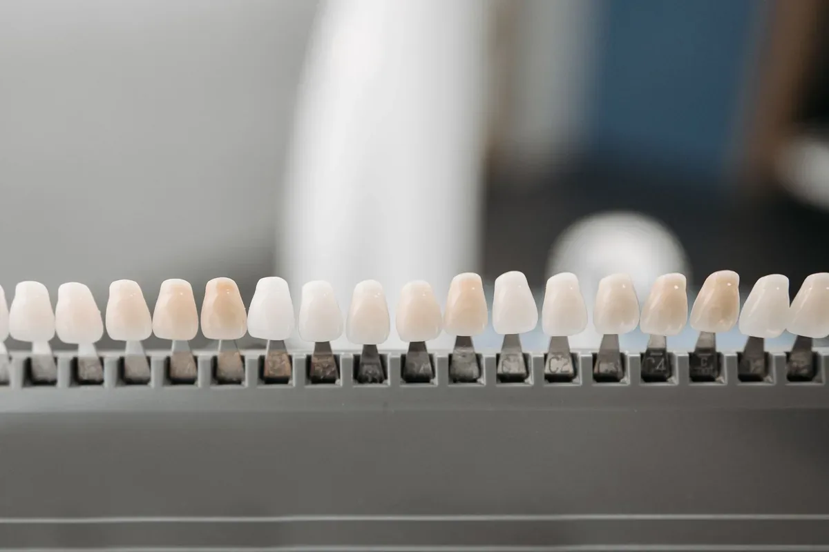
Zirconium Dental Crown: Clinical Indications, Material Structure, and Treatment Protocol
In prosthetic dentistry, when restoring teeth that have lost substance or completing missing teeth with bridge prostheses, the biocompatibility of the material with surrounding tissues is as important as its biomechanical resistance. Metal-supported porcelain crowns, which have long been used as the standard, have various clinical limitations because the metal alloy in the substructure does not allow light to pass through (opaque appearance) and over time can cause gray-purple discoloration at the gum margin. To eliminate these disadvantages, zirconium dioxide-based crowns have taken their place in our clinical practice as a tissue-friendly alternative for the restoration of both anterior and posterior teeth.
Biological and Mechanical Properties of Zirconium
Zirconium used in dentistry is not in its pure mineral form, but rather in the form of yttrium-stabilized tetragonal zirconia polycrystal (Y-TZP). This crystal structure has a phase transformation resistance that stops the progression of microscopic cracks that may occur in the material under chewing forces (900 - 1200 MPa flexural strength).
Another clinically important feature of the material is its low thermal conductivity. Thermal sensitivity to hot and cold foods, which is common in metal-substructure prostheses, is minimized in vital teeth thanks to zirconium’s insulating structure. In addition, the smooth surface of zirconium makes microbial dental plaque retention more difficult. Thanks to its high biocompatibility with gum tissue (gingiva), it is safely used in patients with metal allergies and does not create a gray reflection in the cervical (neck) region even if physiological gum recession occurs over the years.
Clinical Selection: Monolithic and Layered Zirconium
Because every patient’s occlusal dynamics and aesthetic requirements differ, zirconium crowns are planned in two different ways depending on the production technique:
Monolithic Zirconium: Produced from a single zirconium block, with no porcelain layer applied on top. It is the form with the lowest risk of fracture and chipping of the superstructure porcelain. It is especially preferred in posterior (molar) teeth, in cases where chewing forces are intense, in situations with limited vertical bite space, and in patients with bruxism (teeth clenching/grinding) habits.
Layered (Substructure) Zirconium: Obtained by applying feldspathic porcelain that mimics the natural translucency of enamel onto a zirconium substructure that provides durability. Because it offers high light transmission (translucency) and depth perception, it is used in aesthetic restorations of the anterior region.
In Which Cases Is a Zirconium Crown Applied?
The main situations in which we indicate zirconium crowns following clinical examination and radiographic analysis are as follows:
Teeth with coronal substance loss that cannot be restored with composite fillings due to decay or trauma.
Protecting teeth whose brittleness has increased due to fluid loss and tissue weakening after root canal treatment (endodontic treatment) against chewing forces.
Severe internal tooth discoloration caused by antibiotics (tetracycline) or fluorosis, for which whitening (bleaching) does not provide results.
Traditional fixed bridge prostheses used in single or multiple tooth loss and implant-supported crown restorations.
Prosthetic closure of diastema (tooth gap) and minor rotations within clinical limits in patients who do not prefer orthodontic treatment.
Clinical Workflow: Preparation, Digital Impression, and Cementation
The treatment process begins with the related tooth being reduced circumferentially by 1.0 to 1.5 mm under local anesthesia (preparation). To ensure complete seating of the crown at the gum margin and prevent pressure on periodontal tissues, the "chamfer" or rounded shoulder preparation technique is applied.
After the preparation procedure, the gingival sulcus is made visible with retraction cord, and a three-dimensional optical impression is taken using intraoral digital scanners (CAD/CAM). In the same session, acrylic temporary crowns are placed to prevent the patient’s tooth sensitivity and preserve aesthetics. The zirconium crowns, produced and sintered (fired at high temperature) in computer-aided design and milling units in the laboratory, are first tried in the mouth when they arrive at the clinic in terms of marginal fit, contact points with adjacent teeth, and bite height. After fit is approved, the inner surface of the crown is roughened through special procedures and bonded to the tooth using MDP-containing resin cements or resin-modified glass ionomer cements (cementation).
Long-Term Success and Oral Care
The trouble-free service life of zirconium crowns in the mouth depends on the precision of marginal fit and the patient’s oral hygiene habits. The crown itself does not decay; however, bacterial plaque that may leak from the gum margin can lead to secondary decay and gingivitis in the natural tooth tissue beneath the crown. Therefore, brushing twice a day, using special dental flosses (superfloss) or a water flosser for the areas under bridge pontics, and clinical check-ups every six months are essential for maintaining the treatment.