

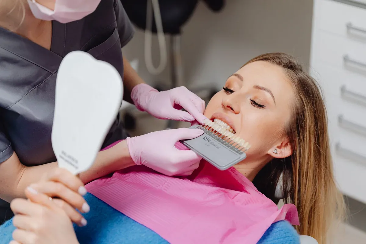
Porcelain Laminate Veneer Applications: Clinical Criteria, Material Structure, and Treatment Process
In prosthetic dentistry, our primary goal is to restore the function and esthetics the patient has lost while preserving healthy tooth structure to the maximum extent possible. While traditional full crown (cap) applications require circumferential reduction of 1.5 to 2 mm from all surfaces of the tooth, porcelain laminate veneer restorations developed in line with the principles of preventive dentistry are a minimally invasive treatment option that preserves healthy enamel tissue.
In the medical literature, this method, referred to as "laminate veneer," involves removing a very thin layer only from the front (labial) surfaces of the teeth that contact the lips and cheeks, or in suitable cases performing no reduction at all, and chemically bonding wafer-thin ceramic restorations to the tooth.
Material Properties: Lithium Disilicate and Feldspathic Ceramics
The clinical success of porcelain laminates and their ability to mimic natural tooth enamel are based on the optical and mechanical properties of the glass ceramic materials used. In clinical practice, two basic types of ceramics are preferred depending on the needs of the case:
Lithium Disilicate Reinforced Ceramics (e.max): These are glass ceramics with increased resistance to chewing forces and flexure (approximately 360 - 400 MPa). They are the material most commonly used in clinical applications because they offer both durability and translucency.
Feldspathic Porcelain: This material is produced directly in the laboratory using a layering technique and has color transitions and optical depth closest to natural tooth enamel. Since its mechanical strength is lower than lithium disilicate, it is preferred in cases with low occlusal stress.
Both materials do not contain a metal substructure, reflect light by refracting it like natural enamel, and show full biological compatibility with gingival tissue. Thanks to their smooth surface, they are resistant to discoloration caused by external factors such as tea, coffee, and tobacco.
Clinical Indications and Contraindications
Long-term success in laminate treatment depends on proper case selection. As clinicians, we determine the indication by analyzing the patient's occlusal relationships and enamel thickness during the intraoral examination.
Indicated Cases (Indications):
Severe intrinsic discoloration caused by antibiotics (tetracycline) or fluorosis that does not respond to tooth whitening (bleaching) protocols.
Closing anatomical spaces called "diastema" between the anterior teeth.
Reconstructing the form and length of teeth with fractured incisal edges or shortened crowns due to trauma or physiological wear.
Prosthetic correction of mild crowding and rotations that do not require orthodontic treatment.
Surface restoration of teeth with structural enamel defects (amelogenesis imperfecta, hypoplasia).
Contraindicated Cases (Contraindications): Porcelain laminates are not a suitable treatment for every patient. In cases with advanced tooth clenching and grinding habits (bruxism), edge-to-edge contact between the upper and lower anterior teeth (tet-a-tet occlusion), or deep crossbite, the risk of ceramic fracture is high. In addition, in patients who do not have sufficient healthy enamel on the tooth surface due to previously placed large fillings or excessive wear, full ceramic or zirconium crowns should be preferred instead of laminates.
Clinical Protocol: Mock-up, Preparation, and Adhesive Cementation
The treatment process begins with taking intraoral impressions of the patient and trying the tooth shapes that will be created at the end of treatment in the patient's mouth beforehand using temporary materials called a "mock-up." At this stage, phonation (the production of sounds during speech), lip support, and occlusal dynamics are directly tested on the teeth.
Following the approved plan, the "preparation" (reduction) stage begins. The basic biomechanical rule of laminate reduction is that the reduction margins must remain entirely within the enamel tissue (average depth of 0.3 to 0.7 mm). The inner (lingual) surfaces of the teeth are not touched.
After the digital impression taken with intraoral scanners, the ceramic laminates are applied to the tooth with the "adhesive cementation" protocol, the most sensitive stage of the treatment. Unlike the adhesives used in traditional crowns, laminates adhere to the tooth through a "bonding" mechanism. The tooth surface is etched with phosphoric acid, while the inner surface of the porcelain is micro-roughened with hydrofluoric acid and silane agents. Then, by using light-cured resin cements, a monoblock (single-piece) chemical integrity is created between the laminate and the tooth enamel. This bond to the enamel allows the porcelain to adhere to the tooth with very high force.
Long-Term Success and Clinical Maintenance
Once the adhesive cementation procedure is completed, porcelain laminates function like natural teeth. However, to preserve the mechanical lifespan of the restorations, patients must avoid parafunctional habits such as nail biting, pen biting, or breaking hard-shelled foods with the front teeth. In patients with a mild tendency to clench their teeth, a protective night guard (occlusal splint) is prepared at the end of treatment.
Since the back surfaces and gingival margins of teeth restored with laminates preserve their natural tissue, brushing twice a day, daily use of dental floss, and routine dental check-ups every 6 months are essential for maintaining both gingival health and the marginal fit of the restoration.