

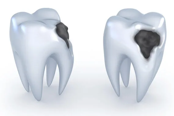
Oral and dental health is one of the cornerstones of bodily integrity and overall quality of life. A healthy digestive system, clear speech, and an aesthetic smile are possible with a complete and healthy set of teeth. However, dietary habits in daily life, genetic predispositions, or deficiencies in oral hygiene can pave the way for decay that threatens the structural integrity of teeth. One of the most fundamental and common preventive/restorative procedures in dentistry, filling treatment enables damaged tooth tissue to be cleaned and restored with biocompatible materials.
If you are experiencing sensitivity, pain, or visible tissue loss in your teeth and are seeking objective, medical, and detailed information about Bursa filling treatment processes, this comprehensive guide will help you understand the treatment steps. This article has been prepared to inform the public in full compliance with healthcare information standards and ethical rules.
How Does Tooth Decay Occur and Why Should It Be Treated?
Tooth decay (caries) is a process that begins when bacteria naturally present in the oral flora break down carbohydrate- and sugar-containing foods and produce acid. Over time, these acids dissolve minerals such as calcium and phosphate in tooth enamel, the hardest tissue in the body, weakening the structure through demineralization. This irreversible erosion, which begins in the enamel layer, progresses toward the dentin layer, which is softer and closer to the nerve network, if not treated.
Once decay reaches the dentin layer, it begins to cause sensitivity to hot, cold, sweet, or sour foods. Before the decayed tissue reaches the vital area at the center of the tooth (the pulp), it should be removed and the resulting space (cavity) filled. A filling procedure not performed in time can allow the infection to reach the tooth’s nerves, making more advanced surgical procedures such as root canal treatment or complete loss of the tooth (extraction) necessary.
In Which Situations Is a Filling Needed?
There is a common belief in society that filling treatment is only applied to black or brown decay lesions. However, in current dentistry approaches, filling materials are also actively used in the repair of many structural problems other than decay:
Physical Trauma: Fracture of a part of the tooth due to a fall, impact, or biting a hard object.
Abrasion and Erosion: Wedge-shaped wear, especially at the gum line, caused by incorrect and forceful tooth brushing habits. Enamel loss due to heavy consumption of acidic drinks.
Teeth Grinding (Bruxism): Cracks and tissue loss on chewing surfaces caused by clenching of the teeth during sleep or under stress.
Replacement of Old Fillings: Replacing old restorations placed years ago that have lost marginal integrity, are leaking, or no longer function properly.
Diastema Closure: Closing the anatomical spaces between front teeth by shaping them with aesthetic filling materials without damaging tooth tissue.
What Are the Dental Filling Materials?
In filling treatments performed in clinics in Bursa, different materials are preferred depending on the patient’s medical needs, the tooth’s position in the mouth, and the chewing forces it is exposed to. The choice of material to be used is determined by the dentist following the clinical examination.
1. Composite Resin Fillings (Aesthetic Fillings)
These are the most commonly preferred restorative materials today, bonding to the tooth with chemical agents and able to closely mimic tooth color. Their durability is enhanced by silica glass particles in their composition. They are placed into the tooth in a paste-like consistency and hardened within seconds using a special light device (halogen or LED), a process called polymerization. They are used to provide both aesthetic and functional integrity in anterior and posterior teeth.
2. Inlay and Onlay Restorations (Porcelain Fillings)
These are preferred when the tissue loss in a tooth is too large to be repaired with a standard composite filling, but too small to require the tooth to be fully prepared and covered. In the clinical setting, the decay is removed and a digital or physical impression of the tooth is taken. In the laboratory, a tooth-compatible piece is prepared from reinforced ceramic (porcelain) materials and fixed to the tooth in a second session with special adhesives. They show outstanding resistance to chewing forces.
3. Glass Ionomer Cements
They are used especially in the treatment of primary teeth, root surface caries, or to create a protective base under composite fillings. They have the property of increasing the tooth’s resistance to decay (cariostatic effect) by gradually releasing the fluoride they contain into the oral environment.
4. Amalgam Fillings
These are traditional metal-alloy fillings containing silver, copper, tin, and mercury. Although they are highly resistant to chewing pressure, their gray/black metallic color does not meet aesthetic expectations. With the development of biocompatible composite and porcelain materials, their clinical use has decreased significantly.
Bursa Filling Treatment: Clinical Application Steps
The process of cleaning a tooth from decay and restoring its function consists of precise steps in accordance with international medical protocols:
1. Examination and Diagnosis: The treatment process begins with radiographic (X-ray) examination and clinical evaluation. The depth of the decay, its proximity to the nerve tissue, and the condition of the surrounding tissues are assessed.
2. Anesthesia Application: To ensure the patient feels no pain or discomfort during the procedure, the relevant tooth and surrounding tissues are numbed with local anesthesia. In superficial enamel wear, anesthesia may not be necessary depending on the dentist’s assessment.
3. Isolation: Completely isolating the tooth to be treated from saliva and breath moisture is critical for the filling to adhere to the tooth (adhesion). For this purpose, cotton rolls or rubber covers called "rubber dam" are used to keep the working area dry.
4. Removal of Decayed Tissue: Using rotary instruments (burs) with special tips, the infected, softened decayed tissue in the enamel and dentin layers is completely removed. Once healthy tooth tissue is reached, the cavity is shaped to retain the filling material.
5. Acid Etching and Application of Bonding Agent: To allow the composite filling to bond chemically to the tooth, the tooth surface is roughened with a very mild acid gel (etching). After the acid is rinsed off, a special bonding resin called "bond" is applied to the tooth and light-cured.
6. Placement of the Material and Curing: The chosen filling material is placed into the cavity in layers (incremental technique). Each layer is cured with a special light source to minimize the risk of material shrinkage. Indentations and protrusions appropriate to the tooth’s anatomy are created.
7. Height Check and Polishing: After the filling procedure is completed, the patient is asked to bite on a special carbon paper. If there are any high or uncomfortable contact points during chewing, they are corrected by grinding them down with burs. In the final stage, the filling surface is smoothed with polishing discs and pastes. A smooth surface makes it harder for bacteria to adhere to that area in the future.
What Should Be Considered After Treatment?
After the clinical procedure is completed, dentists recommend following certain rules to protect oral and dental health:
Effect of Anesthesia: The numbness usually wears off within 2 to 4 hours. During this time, nothing should be chewed, and only warm liquids should be consumed to avoid accidentally biting and injuring the tongue, lip, or cheek tissues.
Sensitivity: During the first few days after the procedure, a mild aching sensation to cold and heat in the tooth is physiologically expected. This sensitivity usually disappears on its own within a few days or weeks as the tissues adapt.
Oral Care Routine: The belief that a filled tooth cannot decay again is mistaken. The junctions where the filling ends and the natural tooth begins can give rise to secondary decay if not cleaned well. Brushing at least twice a day with fluoride toothpaste and the use of dental floss/interdental brushes should not be neglected.
Regular Checkups: Six-month periodic dental checkups should not be skipped for the early detection of restoration margin integrity, possible fractures, or new decay formation.
Preserving the natural tooth structure is always the primary goal from a medical standpoint. A filling treatment performed at the right time and in accordance with scientific protocols is the strongest barrier against tooth loss. Not ignoring symptoms and resolving problems before they worsen through regular dental checkups is the long-term safeguard of your oral and dental health.
Legal Notice and Information
This page content has been prepared solely to inform the public about oral and dental health processes within the framework of the provisions of Law No. 1219 on the Practice of Medicine and its Branches and the Ministry of Health Regulation on Promotional and Informational Activities in Health Services. The information contained herein cannot be used for the diagnosis or treatment of a disease and does not carry a guiding or definitive outcome-promising nature. For an accurate diagnosis and a personalized treatment plan suitable for your oral and dental structure, you must consult a dentist directly and undergo a clinical examination.